AA sequence: Gly-Asp-Cys3-Leu-Pro-His-Leu-Lys-Leu-Cys10-Lys-Glu-Asn-Lys-Asp-Cys16-Cys17-Ser-Lys-Lys-Cys21-Lys-Arg-Arg-Gly-Thr-Asn-Ile-Glu-Lys-Arg-Cys32-Arg-OH
Disulfide bonds: Cys3-Cys17, Cys10-Cys21 and Cys16-Cys32
Length (aa): 33
Formula: C156H260N56O46S6
Molecular Weight: 3858.8 Da
Appearance: White lyophilized solid
Solubility: water and saline buffer
CAS number: not available
Source: Synthetic
Purity rate: > 98 %
Maurocalcine
170 $ – 850 $
Maurocalcine, a potent agonist of ryanodine receptors
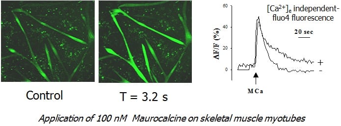
Maurocalcine, a component of the venom of Scorpio maurus palmatus, was discovered by Michel De Waard, co-founder of Smartox. Maurocalcine folds according to an inhibitor cysteine knot. Such as Imperatoxin A, Maurocalcine acts as a high affinity agonist of the type-1 ryanodine receptor expressed in skeletal muscles with an affinity in the 10 nM range. Maurocalcine induces an increase in channel opening probability accompanied by sudden transitions to long lasting subconductance states. Maurocalcine has also been characterized as a cell penetrating peptide and its pharmacological activity can be observed upon extracellular perfusion.
Redox-sensitive stimulation of type-1 ryanodine receptors by the scorpion toxin maurocalcine. Cell Calcium
The scorpion toxin maurocalcine acts as a high affinity agonist of the type-1 ryanodine receptor expressed in skeletal muscle. Here, we investigated the effects of the reducing agent dithiothreitol or the oxidizing reagent thimerosal on type-1 ryanodine receptor stimulation by maurocalcine. Maurocalcine addition to sarcoplasmic reticulum vesicles actively loaded with calcium elicited Ca²⁺ release from native vesicles and from vesicles pre-incubated with dithiothreitol; thimerosal addition to native vesicles after Ca²⁺ uptake completion prevented this response. Maurocalcine enhanced equilibrium [³H]-ryanodine binding to native and to dithiothreitol-treated reticulum vesicles, and increased 5-fold the apparent Ki for Mg²⁺ inhibition of [³H]-ryanodine binding to native vesicles. Single calcium release channels incorporated in planar lipid bilayers displayed a long-lived open sub-conductance state after maurocalcine addition. The fractional time spent in this sub-conductance state decreased when lowering cytoplasmic [Ca²⁺] from 10 μM to 0.1 μM or at cytoplasmic [Mg²⁺]≥30 μM. At 0.1 μM [Ca²⁺], only channels that displayed poor activation by Ca²⁺ were readily activated by 5 nM maurocalcine; subsequent incubation with thimerosal abolished the sub-conductance state induced by maurocalcine. We interpret these results as an indication that maurocalcine acts as a more effective type-1 ryanodine receptor channel agonist under reducing conditions.
Ronjat M, et al. (2013) Redox-sensitive stimulation of type-1 ryanodine receptors by the scorpion toxin maurocalcine. Cell Calcium. PMID: 23623374
Maurocalcine interacts with the cardiac ryanodine receptor without inducing channel modification
We have previously shown that MCa (maurocalcine), a toxin from the venom of the scorpion Maurus palmatus, binds to RyR1 (type 1 ryanodine receptor) and induces strong modifications of its gating behaviour. In the present study, we investigated the ability of MCa to bind to and modify the gating process of cardiac RyR2. By performing pull-down experiments we show that MCa interacts directly with RyR2 with an apparent affinity of 150 nM. By expressing different domains of RyR2 in vitro, we show that MCa binds to two domains of RyR2, which are homologous with those previously identified on RyR1. The effect of MCa binding to RyR2 was then evaluated by three different approaches: (i) [(3)H]ryanodine binding experiments, showing a very weak effect of MCa (up to 1 muM), (ii) Ca(2+) release measurements from cardiac sarcoplasmic reticulum vesicles, showing that MCa up to 1 muM is unable to induce Ca(2+) release, and (iii) single-channel recordings, showing that MCa has no effect on the open probability or on the RyR2 channel conductance level. Long-lasting opening events of RyR2 were observed in the presence of MCa only when the ionic current direction was opposite to the physiological direction, i.e. from the cytoplasmic face of RyR2 to its luminal face. Therefore, despite the conserved MCa binding ability of RyR1 and RyR2, functional studies show that, in contrast with what is observed with RyR1, MCa does not affect the gating properties of RyR2. These results highlight a different role of the MCa-binding domains in the gating process of RyR1 and RyR2.
Altafaj X, et al. (2007) Maurocalcine interacts with the cardiac ryanodine receptor without inducing channel modification. Biochem J. PMID: 17537000
Chemical synthesis and characterization of maurocalcine, a scorpion toxin that activates Ca(2+) release channel/ryanodine receptors
Maurocalcine is a novel toxin isolated from the venom of the chactid scorpion Scorpio maurus palmatus. It is a 33-mer basic peptide cross-linked by three disulfide bridges, which shares 82% sequence identity with imperatoxin A, a scorpion toxin from the venom of Pandinus imperator. Maurocalcine is peculiar in terms of structural properties since it does not possess any consensus motif reported so far in other scorpion toxins. Due to its low concentration in venom (0.5% of the proteins), maurocalcine was chemically synthesized by means of an optimized solid-phase method, and purified after folding/oxidation by using both C18 reversed-phase and ion exchange high-pressure liquid chromatographies. The synthetic product (sMCa) was characterized. The half-cystine pairing pattern of sMCa was identified by enzyme-based cleavage and Edman sequencing. The pairings were Cys3-Cys17, Cys10-Cys21, and Cys16-Cys32. In vivo, the sMCa was lethal to mice following intracerebroventricular inoculation (LD(50), 20 microg/mouse). In vitro, electrophysiological experiments based on recordings of single channels incorporated into planar lipid bilayers showed that sMCa potently and reversibly modifies channel gating behavior of the type 1 ryanodine receptor by inducing prominent subconductance behavior.
Fajloun, Z., et al. (2000) Chemical synthesis and characterization of maurocalcine, a scorpion toxin that activates Ca(2+) release channel/ryanodine receptors, FEBS Lett. PMID: 10713267
A new fold in the scorpion toxin family, associated with an activity on a ryanodine-sensitive calcium channel
We determined the structure in solution by (1)H two-dimensional NMR of Maurocalcine from the venom of Scorpio maurus. This toxin has been demonstrated to be a potent effector of ryanodyne-sensitive calcium channel from skeletal muscles. This is the first description of a scorpion toxin which folds following the Inhibitor Cystine Knot fold (ICK) already described for numerous toxic and inhibitory peptides, as well as for various protease inhibitors. Its three dimensional structure consists of a compact disulfide-bonded core from which emerge loops and the N-terminus. A double-stranded antiparallel beta-sheet comprises residues 20-23 and 30-33. A third extended strand (residues 9-11) is perpendicular to the beta-sheet. Maurocalcine structure mimics the activating segment of the dihydropyridine receptor II-III loop and is therefore potentially useful for dihydropyridine receptor/ryanodine receptor interaction studies.
Mosbah, A., et al. (2000) A new fold in the scorpion toxin family, associated with an activity on a ryanodine-sensitive calcium channel, Proteins. PMID: 10861934